One of the most advanced imaging technologies for studying biological molecules—cryo-electron microscopy (Cryo-EM), often described as a “revolution” in structural biology—is about to have its first specialized laboratory in Southeastern Europe. This groundbreaking facility will be located at the National Hellenic Research Foundation (NHRF) in Athens, Greece.
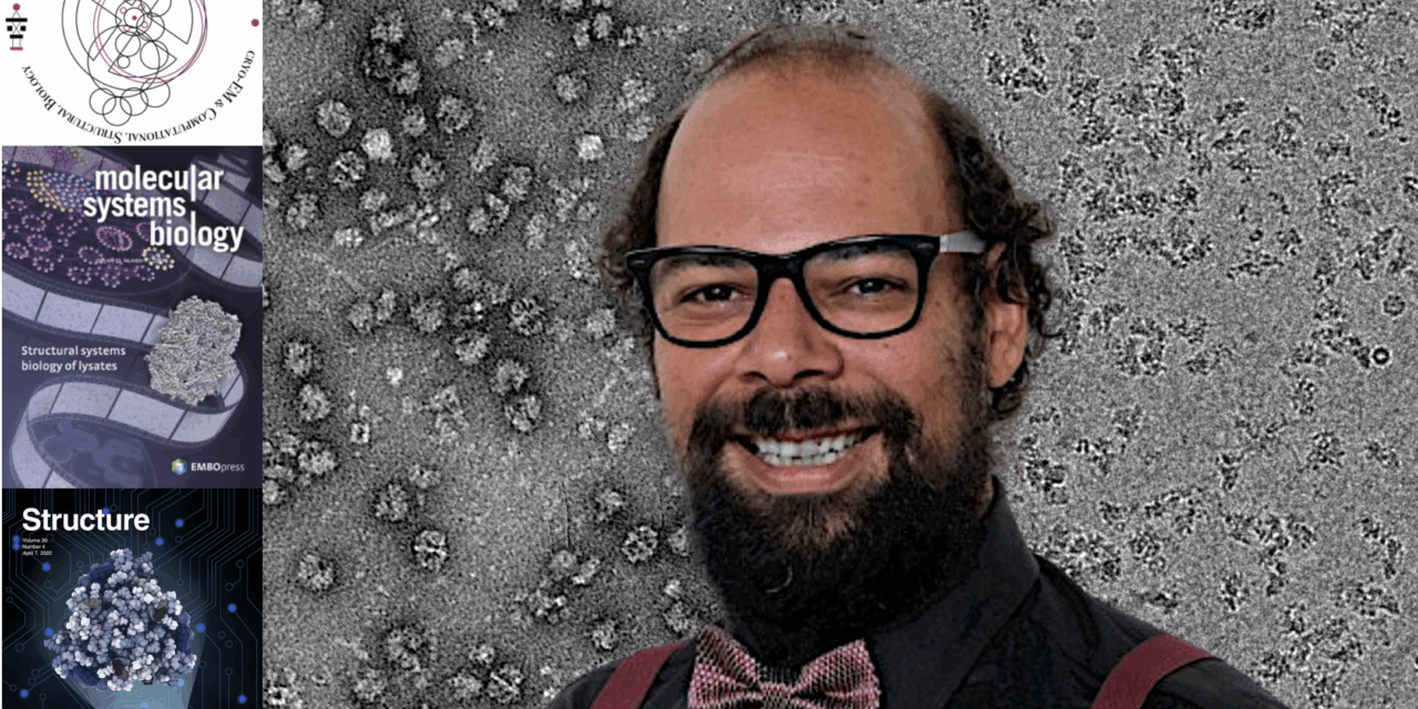
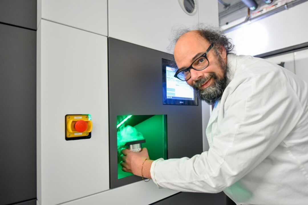
At the helm of this ambitious initiative is Professor Panagiotis Kastritis, a distinguished Greek researcher with an international career, currently serving in the Department of Biochemistry and Biotechnology at Halle-Wittenberg University in Germany.
Cryo-electron microscopy—whose developers, Jacques Dubochet, Joachim Frank and Richard Henderson were jointly awarded the 2017 Nobel Prize in Chemistry—is revolutionizing how scientists observe biological molecules. Previously, viewing proteins and their complex mechanisms at the atomic level posed significant challenges. Cryo-EM allows researchers to flash-freeze biological samples in liquid nitrogen, preserving them for high-resolution imaging using electromagnetic lenses. Advanced software then processes these images into detailed 3D molecular structures, enabling an unprecedented understanding of how cells function.
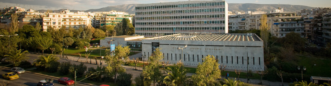
The new lab will be equipped with four state-of-the-art Cryo-EM microscopes: the Krios G4, Aquilos 2, Talos L120C (S)TEM, and Spectra 200 (S)TEM. Procurement contracts for these instruments have already been signed, and renovation of the facility space is underway. This Cryo-EM laboratory will become an integral part of the new Theranostics Center of Excellence, a research and innovation hub at the NHRF focused on cancer diagnostics and therapeutics, funded by the European Recovery and Resilience Facility.
Professor Kastritis: From Greece to Germany and back again
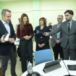
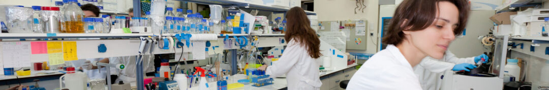
Professor Kastritis, recipient of EU ERA Chair funding, is leading the lab’s development at the NHRF. He has already assembled a multidisciplinary team that is being trained in Cryo-EM methodologies at his lab in Germany. This team is preparing to manage the entire spectrum of sample preparation, imaging, and analysis. Beyond technical training, Kastritis is also transferring operational expertise to Greece with the goal of establishing international collaborations. “Extroversion is absolutely critical for such an expensive and valuable technology coming to Greece,” he emphasizes.
Professor Kastritis began his journey into the microscopic world of cells with a biology degree from the National and Kapodistrian University of Athens. It was during a Molecular Biophysics course taught by Professor Stavros Hamodrakas—a pioneer in computational biology—that he first became captivated by structural biology, a field striving to understand cellular functions through analytical methods. Ηe recalls: “It’s astonishing that we’re made up of billions of cells that communicate with each other. Biophysics helped me see how physical laws explain how cells work. This mathematical modeling of biology fascinated me.”
Until Cryo-EM’s rise, the primary tools for structural biology, which looks at proteins and nucleic acids at a molecular level, were X-ray crystallography and NMR spectroscopy. However, Cryo-EM allows scientists to observe complex biomolecules in unprecedented detail—particularly those resistant to analysis by older methods. This has opened new pathways toward understanding cellular processes at their most fundamental level.
Although known since the 1960s, Cryo-EM only recently surged in relevance. “In recent years, everything changed,” says Kastritis. “Microscopes became more stable, cameras more advanced, graphics cards and data analysis software improved drastically. Sample preparation became semi-automated. That’s why the Nobel Prize was awarded in 2017 to the scientists who made this method what it is today.”
In 2018, Kastritis established the Cryo-EM lab at Halle-Wittenberg University, where he has managed multimillion-euro research funding and published studies in top-tier scientific journals. Since 2023, he has also served as a visiting researcher at the NHRF, and received a €2.5 million ERA Chair grant to bring Cryo-EM expertise to Greece. Through this project, he also hopes to strengthen the exchange between the NHRF and research groups in Halle-Wittenberg University.
Reflecting on this scientific journey, he says: “When you’re a student, the cell’s complexity feels incomprehensible. Even now, I don’t fully understand its entirety. While equations and experiments help us analyze some aspects, the cell remains a profound mystery. But now we can systematically approach this complexity more than ever before. Cryo-EM has truly transformed our understanding of biological macromolecules and their behavior.”
So what is the ultimate purpose of this expanding understanding? “Cryo-EM is a completely new way of trying to understand how cells work. Through my research, I break open cells to examine their contents—to see their structures and understand what’s inside. Each image we obtain reveals new knowledge about cellular metabolism. I want to understand the architecture of life itself. And through collaboration with medical and biotech experts, we aim to answer critical questions surrounding cancer, Alzheimer’s, and other diseases,” he concludes.
Edited and translated to English from the original APE-MPA article by Maria Kouzinopoulou.
Read more from Greek News Agenda:
TAGS: BIOCHEMISTRY | INNOVATION | RESEARCH | SCIENCE & TECHNOLOGY